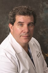
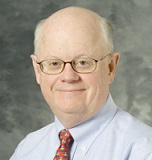
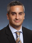
What are the most exciting strides forward in GI/endoscopy technology? Ten gastroenterologists weigh in on what they think are the biggest advancements in the field.
Ask a Gastroenterologist is a weekly series of questions posed to GI physicians around the country on business and clinical issues affecting the field of gastroenterology. We invite all gastroenterologists to submit responses. Next week's question: Does it make sense for gastroenterologists to join accountable care organizations?
Please submit responses to Carrie Pallardy at cpallardy@beckershealthcare.com by Thursday, Oct. 2, at 5 p.m. CST.
 Deepak Agrawal, MD, Assistant Professor of Internal Medicine at UT Southwestern Medical Center (Dallas): Gastrointestinal endoscopy has seen some exciting improvements and innovations in the last few years. They are a product of better technology, better equipment and bold, innovative uses of the improved technology. One of the most important and widely embraced improvements is in endoscopic image clarity. The high resolution, high definition and high magnification endoscopy now allow us to examine the mucosa and even overlying capillaries in very fine details. Most endoscopes now have narrow band imaging wherein a light of very narrow wavelength is projected on the mucosa. This special light can help the endoscopist differentiate lesions as benign or malignant. The image quality is like watching super bowl on an ultra high def TV; it would be very difficult to go back to a normal screen again. Special endoscopes now even allow endoscopists to see individual cells. This technique is called confocal endoscopy.
The other exciting innovation is improvement in our ability to close holes or defects in the wall of the gut. This has been made possible due to special large clips such as over the scope clips or endoscopic suturing devices. This has increased the confidence of the endoscopists to attempt removing large polyps or superficial tumors through techniques such as endoscopic mucosal resection or endoscopic submucosal dissection. These procedures have helped many patients avoid surgery.
Maxwell Chait MD, FACP, FACG, FASGE, AGAF, Columbia Doctors Medical Group (Hartsdale, N.Y.): The field of gastrointestinal endoscopy has been burgeoning with emerging new and exciting technology in the past few years. Advances in endoscopy visualization include illumination with narrow-band imaging, florescence endoscopy, chromoendoscopy confocal endoscopy and the nearly 360 degree view Fuse endoscope. Endoscopic ultrasound provides assessment, tissue and processes for drainage not previously considered. Endoscopic biopsy including CLO test has allowed treatment and eradication of Helicobacter pylori infection and thus treatment of gastritis, peptic ulcer and gastric MALT lymphoma and prevention of gastric cancer.
There have been significant treatment modalities developed, many of which continue to evolve, that broaden the possibilities of treatment with success that rival surgical therapies. Radiofrequency ablation for high-grade dysplasia in Barrett's esophagus is available for prevention of esophageal cancer; endoscopic mucosal resection and endoscopic submucosal dissection for treatment of Barrett's esophagus and superficial cancers of the esophagus, stomach and colon. Natural Orifice Transluminal Endoscopic Surgery offers the possibility of removing organs such as the gallbladder or appendix via natural orifices such as the mouth and results in scarless surgery. Peroral endoscopic myotomy provides a natural orifice approach to performing the Heller myotomy for the treatment of achalasia. Endoscopic full thickness resection is being evaluated to allow complete removal of tumors without any scars.
 Ashkan Farhadi, MD,Orange Coast Memorial Medical Center (Fountain Valley, Calif.): It is very exciting to practice gastroenterology in an era when we have the capability to investigate the gastrointestinal lumen with high definition cameras. Now, we can utilize our Mars Rover equivalent called video capsule endoscopy in which a capsule takes films as it travels through the GI tract and sends the images to a receiver outside of body. We can also implant a wireless "laboratory station" in the esophagus to sample and measure the acid exposure.
The capability also exists to do ultrasound from the inside of the gut lumen and sample tissues outside of our GI tract such as those found in the lung or pancreas through endoscopy with guidance of ultrasound. Corrective surgeries for hiatal hernias can be done endoscopically using suturing devices that pass through the scope. We may also see NOTES (natural orifice transluminal endoscopic surgery) which are a whole array of surgeries being done by special endoscopes that pass through GI tract and travel beyond the GI tract boundaries by cutting a passage through it. This allows formerly invasive surgery such as an appendectomy or cholecystectomy to be performed without any skin incision.
 Jeffrey Fine, MD, Chief of Gastroenterology, Medical and Surgical Clinic of Irving, gastroenterologist, Star Medical Center (Plano, Texas) Las Colinas Medical Center (Irving,Texas): It's an exciting time for the field. During the past few years, there have been many advances in gastroenterology. Most notably, a new treatment for hepatitis C genotype 2, a new treatment for Crohn's disease using the medicines UCERIS and Entyvio and high resolution and esophageal manometery, a procedure that helps determine how the muscle of the esophagus and the sphincter works by measuring the pressures they generate.
William Katkov, MD, Providence Saint John's Health Center, Santa Monica, Calif.:Technological innovation has been a cornerstone of gastroenterology and its advances over the past several decades. In recent years, strides have been made in imaging techniques of the GI tract. In the realm of intestinal disease and the diagnosis of GI bleeding of obscure origin, capsule endoscopy is a major step forward and a part of daily GI clinical practice. Enhanced visualization of colon adenomas has been made possible by several developments to improve detection rates including narrow band imaging (NBI) and high-definition colonoscopy. An additional exciting technology is transient elastography which provides a reliable non-invasive way to assess the degree of liver fibrosis.
Michael Kochman, MD, AGAF, chair, AGA Center for GI Innovation and Technology: Innovation in gastroenterology is not dead and is indeed picking up the pace. As a minimally invasive, and mostly ambulatory specialty, we play a large role in the provision of quality high-value care by both cognitive and procedural interactions with our patients. Over the past few years there have been significant technological innovations surrounding endoscopic procedures, some of which are now FDA-approved, and some likely to be approved in the near future.
A major area of clinical and research interest continues to be colonic neoplasia detection. Two innovations over the past year are likely to benefit patients and may also prove to be cost-effective. The Covidien PillCam Colon capsule is now approved for patients who underwent a failed colonoscopy due to technical reasons and the Ethicon Sedasys system affords an additional way to administer propofol. Two other major therapeutic areas of interest that are compelling are transoral procedures for reflux and obesity. Both Medigus and EGS have FDA approval for transoral reflux systems and at least two transoral obesity devices are awaiting FDA approval decisions with others in clinical trial in the U.S.
The AGA Center for GI Innovation and Technology supports innovation and the development of new technology in gastroenterology, hepatology, nutrition and obesity by guiding medical device and therapeutics innovators through the technology development and adoption process. Through the center, we hope to continue to advance new technologies in GI.
 Mark Reichelderfer, MD, Professor, Gastroenterology and Hepatology, University of Wisconsin School of Medicine and Public Health (Madison): One of the most exciting advances in the gastroenterology field took place in August of this year when the FDA approved Cologuard, the first and only noninvasive stool DNA screening test for colorectal cancer. The test detects both DNA and blood biomarkers associated with colorectal cancer and pre-cancer in the stool and is highly accurate. In the 10,000-patient DeeP-C study published in the New England Journal of Medicine, the sensitivity of Cologuard in detecting patients with colorectal cancer was 92 percent, versus 74 percent for existing stool-based screening methods. Cologuard also detected 42 percent of precancerous lesions and achieved 87 percent specificity.
Colorectal cancer is a highly preventable cancer if caught early, which is why increasing screening compliance rates is crucial. Colonoscopy remains the gold standard in colon cancer screening, but I think Cologuard is a new option that will appeal to those patients who are unable to undergo colonoscopy or who are wary of more invasive screening options. Cologuard does not require the same dietary restriction or bowel prep associated with other screening tests and the patient can take the test in the privacy of their own home, then mail the sample back to the lab for testing. Cologuard's accuracy and patient-friendly design should work favorably together to increase screening compliance rates, which is our ultimate goal as gastroenterologists.
Patrick Takahashi, MD, CMIO and Chief of Gastroenterology Section of St. Vincent Medical Center (Los Angeles): Some of the more exciting advances in the gastrointestinal field include equipment improvements. We now have a company focusing upon improving our ability to visualize the colon in a more complete fashion. The ability to see the colon at different angles and perspectives is quite an improvement, leaving less chance of a missed lesion. Improvements in the ability to visualize abnormal tissue through various filters in the scope have also helped to target abnormal lesions that need to be either removed, biopsied, fulgurated or monitored. This Narrow Beam Imaging has helped tremendously in looking at Barrett's esophagus as well as identifying difficult to see sessile polyps within the colon to name a few examples.
ERCP catheters and devices have also improved, providing a much smoother experience for both endoscopist and patient. Cannulation percentages will continue to improve with decreasing risks of complications such as pancreatitis as we continue to learn about the pancreaticobiliary tract. The ability to extract stones has never been easier than now as we have newer disposable baskets and the like which help us accomplish our goals. The field of gastroenterology continues to improve, and technology will lead the way, enhancing the experience for all involved.
 Pankaj Vashi, MD, Lead National Medical Director, National Clinical Director of Gastroenterology/Nutrition, Metabolic Support and Gastroenterology, Cancer Treatment Centers of America at Midwestern Regional Medical Center (Zion, Ill.): In the past few years, there have been many advances in GI technology. Five are worth mentioning specifically:
1. Confocal laser endo-microscope helps identify premalignant and malignant cells in real time when we are doing a procedure. It is good for patients with Barrett's epithelium, pancreatic cysts, biliary strictures and early gastric cancers.
2. Stool DNA testing as a supporting test to detect colon cancer along with screening colonoscopy. It has very high sensitivity.
3. Endoscopic radio frequency ablation for early esophageal cancers along with endoscopic mucosal resections.
4. Endoscopic suturing devices for prevention of migration on endoscopes and closure of enteral fistulas and small perforations. This has a steep learning curve.
5. Single and double balloon enteroscopy for small bowel visualization.
We also look toward the future for even more new technologies like endoscopic drug delivery systems for pancreatic tumors and digital spyglass technology which will allow us to visualize the biliary tree and do biopsies. Neither are yet approved by the FDA
Jay N. Yepuri, MD, Digestive Health Associates of Texas (Bedford): Over the past few years the most exciting advances in GI technology have been in the realm of therapeutic endoscopy. In the esophagus, the ability to definitively treat Barrett's Esophagus with radiofrequency ablation (HALO) and endoscopic mucosal resection have enabled a paradigm shift in the management of both this condition and early, superficial esophageal adenocarcinoma.
POEM (per oral endoscopic myotomy) shows real promise for the management of achalasia. The management of GERD will change significantly with the introduction of Stretta — a purely endoscopic platform that uses RF energy to enhance the integrity of the lower esophageal sphincter. In the stomach, EMR and endoscopic submucosal dissection have allowed for definitive endoscopic management of early, superficial gastric cancers.
In the pancreaticobiliary arena, advances in EUS have allowed for this modality to transform from a purely diagnostic technique to one with true therapeutic potential. Rendezvous/access procedures and alcohol ablation of cystic pancreatic lesions are two examples of this evolution. Spyglass technology has made choledochoscopy during ERCP more straightforward and significantly broadened its clinical utility/role. The management of complex biliary stone disease and the evaluation of indeterminate biliary strictures have been transformed by this technology. Finally, the advent of single/double balloon enteroscopy in conjunction with advances in capsule technology (by both Medivators and Given Imaging) have radically advanced our ability to image and intervene in the small intestine."
More articles on gastroenterology:
7 gastroenterologists in the news
Are current endoscope cleaning protocols enough?
6 things to know about colonoscopy cost & what a reimbursement cut would mean
Ask a Gastroenterologist is a weekly series of questions posed to GI physicians around the country on business and clinical issues affecting the field of gastroenterology. We invite all gastroenterologists to submit responses. Next week's question: Does it make sense for gastroenterologists to join accountable care organizations?
Please submit responses to Carrie Pallardy at cpallardy@beckershealthcare.com by Thursday, Oct. 2, at 5 p.m. CST.
 Deepak Agrawal, MD, Assistant Professor of Internal Medicine at UT Southwestern Medical Center (Dallas): Gastrointestinal endoscopy has seen some exciting improvements and innovations in the last few years. They are a product of better technology, better equipment and bold, innovative uses of the improved technology. One of the most important and widely embraced improvements is in endoscopic image clarity. The high resolution, high definition and high magnification endoscopy now allow us to examine the mucosa and even overlying capillaries in very fine details. Most endoscopes now have narrow band imaging wherein a light of very narrow wavelength is projected on the mucosa. This special light can help the endoscopist differentiate lesions as benign or malignant. The image quality is like watching super bowl on an ultra high def TV; it would be very difficult to go back to a normal screen again. Special endoscopes now even allow endoscopists to see individual cells. This technique is called confocal endoscopy.
 The other exciting innovation is improvement in our ability to close holes or defects in the wall of the gut. This has been made possible due to special large clips such as over the scope clips or endoscopic suturing devices. This has increased the confidence of the endoscopists to attempt removing large polyps or superficial tumors through techniques such as endoscopic mucosal resection or endoscopic submucosal dissection. These procedures have helped many patients avoid surgery.
Maxwell Chait MD, FACP, FACG, FASGE, AGAF, Columbia Doctors Medical Group (Hartsdale, N.Y.): The field of gastrointestinal endoscopy has been burgeoning with emerging new and
 exciting technology in the past few years. Advances in endoscopy visualization include illumination with narrow-band imaging, florescence endoscopy, chromoendoscopy confocal endoscopy and the nearly 360 degree view Fuse endoscope. Endoscopic ultrasound provides assessment, tissue and processes for drainage not previously considered. Endoscopic biopsy including CLO test has allowed treatment and eradication of Helicobacter pylori infection and thus treatment of gastritis, peptic ulcer and gastric MALT lymphoma and prevention of gastric cancer.
 There have been significant treatment modalities developed, many of which continue to evolve, that broaden the possibilities of treatment with success that rival surgical therapies. Radiofrequency ablation for high-grade dysplasia in Barrett's esophagus is available for prevention of esophageal cancer; endoscopic mucosal resection and endoscopic submucosal dissection for treatment of Barrett's esophagus and superficial cancers of the esophagus, stomach and colon. Natural Orifice Transluminal Endoscopic Surgery offers the possibility of removing organs such as the gallbladder or appendix via natural orifices such as the mouth and results in scarless surgery. Peroral endoscopic myotomy provides a natural orifice approach to performing the Heller myotomy for the treatment of achalasia. Endoscopic full thickness resection is being evaluated to allow complete removal of tumors without any scars.
 Ashkan Farhadi, MD,Orange Coast Memorial Medical Center (Fountain Valley, Calif.): It is very exciting to practice gastroenterology in an era when we have the capability to investigate the gastrointestinal lumen with high definition cameras. Now, we can utilize our Mars Rover equivalent called video capsule endoscopy in which a capsule takes films as it travels through the GI tract and sends the images to a receiver outside of body. We can also implant a wireless "laboratory station" in the esophagus to sample and measure the acid exposure.
 The capability also exists to do ultrasound from the inside of the gut lumen and sample tissues outside of our GI tract such as those found in the lung or pancreas through endoscopy with guidance of ultrasound. Corrective surgeries for hiatal hernias can be done endoscopically using suturing devices that pass through the scope. We may also see NOTES (natural orifice transluminal endoscopic surgery) which are a whole array of surgeries being done by special endoscopes that pass through GI tract and travel beyond the GI tract boundaries by cutting a passage through it. This allows formerly invasive surgery such as an appendectomy or cholecystectomy to be performed without any skin incision.
 Jeffrey Fine, MD, Chief of Gastroenterology, Medical and Surgical Clinic of Irving, gastroenterologist, Star Medical Center (Plano, Texas) Las Colinas Medical Center (Irving,Texas): It's an exciting time for the field. During the past few years, there have been many advances in gastroenterology. Most notably, a new treatment for hepatitis C genotype 2, a new treatment for Crohn's disease using the medicines UCERIS and Entyvio and high resolution and esophageal manometery, a procedure that helps determine how the muscle of the esophagus and the sphincter works by measuring the pressures they generate.
William Katkov, MD, Providence Saint John's Health Center, Santa Monica, Calif.:Technological
 innovation has been a cornerstone of gastroenterology and its advances over the past several decades. In recent years, strides have been made in imaging techniques of the GI tract. In the realm of intestinal disease and the diagnosis of GI bleeding of obscure origin, capsule endoscopy is a major step forward and a part of daily GI clinical practice. Enhanced visualization of colon adenomas has been made possible by several developments to improve detection rates including narrow band imaging (NBI) and high-definition colonoscopy. An additional exciting technology is transient elastography which provides a reliable non-invasive way to assess the degree of liver fibrosis.
Michael Kochman, MD, AGAF, chair, AGA Center for GI Innovation and Technology: Innovation in gastroenterology is not dead and is indeed picking up the pace. As a
 minimally invasive, and mostly ambulatory specialty, we play a large role in the provision of quality high-value care by both cognitive and procedural interactions with our patients. Over the past few years there have been significant technological innovations surrounding endoscopic procedures, some of which are now FDA-approved, and some likely to be approved in the near future.
A major area of clinical and research interest continues to be colonic neoplasia detection. Two innovations over the past year are likely to benefit patients and may also prove to be cost-effective. The Covidien PillCam Colon capsule is now approved for patients who underwent a failed colonoscopy due to technical reasons and the Ethicon Sedasys system affords an additional way to administer propofol. Two other major therapeutic areas of interest that are compelling are transoral procedures for reflux and obesity. Both Medigus and EGS have FDA approval for transoral reflux systems and at least two transoral obesity devices are awaiting FDA approval decisions with others in clinical trial in the U.S.
The AGA Center for GI Innovation and Technology supports innovation and the development of new technology in gastroenterology, hepatology, nutrition and obesity by guiding medical device and therapeutics innovators through the technology development and adoption process. Through the center, we hope to continue to advance new technologies in GI.
 Mark Reichelderfer, MD, Professor, Gastroenterology and Hepatology, University of Wisconsin School of Medicine and Public Health (Madison): One of the most exciting advances in the gastroenterology field took place in August of this year when the FDA approved Cologuard, the first and only noninvasive stool DNA screening test for colorectal cancer. The test detects both DNA and blood biomarkers associated with colorectal cancer and pre-cancer in the stool and is highly accurate. In the 10,000-patient DeeP-C study published in the New England Journal of Medicine, the sensitivity of Cologuard in detecting patients with colorectal cancer was 92 percent, versus 74 percent for existing stool-based screening methods. Cologuard also detected 42 percent of precancerous lesions and achieved 87 percent specificity.
Colorectal cancer is a highly preventable cancer if caught early, which is why increasing screening compliance rates is crucial. Colonoscopy remains the gold standard in colon cancer screening, but I think Cologuard is a new option that will appeal to those patients who are unable to undergo colonoscopy or who are wary of more invasive screening options. Cologuard does not require the same dietary restriction or bowel prep associated with other screening tests and the patient can take the test in the privacy of their own home, then mail the sample back to the lab for testing. Cologuard's accuracy and patient-friendly design should work favorably together to increase screening compliance rates, which is our ultimate goal as gastroenterologists.
Patrick Takahashi, MD, CMIO and Chief of Gastroenterology Section of St. Vincent Medical Center (Los Angeles): Some of the more exciting advances in the gastrointestinal field
 include equipment improvements. We now have a company focusing upon improving our ability to visualize the colon in a more complete fashion. The ability to see the colon at different angles and perspectives is quite an improvement, leaving less chance of a missed lesion. Improvements in the ability to visualize abnormal tissue through various filters in the scope have also helped to target abnormal lesions that need to be either removed, biopsied, fulgurated or monitored. This Narrow Beam Imaging has helped tremendously in looking at Barrett's esophagus as well as identifying difficult to see sessile polyps within the colon to name a few examples.
 ERCP catheters and devices have also improved, providing a much smoother experience for both endoscopist and patient. Cannulation percentages will continue to improve with decreasing risks of complications such as pancreatitis as we continue to learn about the pancreaticobiliary tract. The ability to extract stones has never been easier than now as we have newer disposable baskets and the like which help us accomplish our goals. The field of gastroenterology continues to improve, and technology will lead the way, enhancing the experience for all involved.
 Pankaj Vashi, MD, Lead National Medical Director, National Clinical Director of Gastroenterology/Nutrition, Metabolic Support and Gastroenterology, Cancer Treatment Centers of America at Midwestern Regional Medical Center (Zion, Ill.): In the past few years, there have been many advances in GI technology. Five are worth mentioning specifically:
1. Confocal laser endo-microscope helps identify premalignant and malignant cells in real time when we are doing a procedure. It is good for patients with Barrett's epithelium, pancreatic cysts, biliary strictures and early gastric cancers.
2. Stool DNA testing as a supporting test to detect colon cancer along with screening colonoscopy. It has very high sensitivity.
3. Endoscopic radio frequency ablation for early esophageal cancers along with endoscopic mucosal resections.
4. Endoscopic suturing devices for prevention of migration on endoscopes and closure of enteral fistulas and small perforations. This has a steep learning curve.
5. Single and double balloon enteroscopy for small bowel visualization.
We also look toward the future for even more new technologies like endoscopic drug delivery systems for pancreatic tumors and digital spyglass technology which will allow us to visualize the biliary tree and do biopsies. Neither are yet approved by the FDA
Jay N. Yepuri, MD, Digestive Health Associates of Texas (Bedford): Over the past few years the most exciting advances in GI technology have been in the realm of therapeutic
 endoscopy. In the esophagus, the ability to definitively treat Barrett's Esophagus with radiofrequency ablation (HALO) and endoscopic mucosal resection have enabled a paradigm shift in the management of both this condition and early, superficial esophageal adenocarcinoma.
 POEM (per oral endoscopic myotomy) shows real promise for the management of achalasia. The management of GERD will change significantly with the introduction of Stretta — a purely endoscopic platform that uses RF energy to enhance the integrity of the lower esophageal sphincter. In the stomach, EMR and endoscopic submucosal dissection have allowed for definitive endoscopic management of early, superficial gastric cancers.
In the pancreaticobiliary arena, advances in EUS have allowed for this modality to transform from a purely diagnostic technique to one with true therapeutic potential. Rendezvous/access procedures and alcohol ablation of cystic pancreatic lesions are two examples of this evolution. Spyglass technology has made choledochoscopy during ERCP more straightforward and significantly broadened its clinical utility/role. The management of complex biliary stone disease and the evaluation of indeterminate biliary strictures have been transformed by this technology. Finally, the advent of single/double balloon enteroscopy in conjunction with advances in capsule technology (by both Medivators and Given Imaging) have radically advanced our ability to image and intervene in the small intestine."
More articles on gastroenterology:
7 gastroenterologists in the news
Are current endoscope cleaning protocols enough?
6 things to know about colonoscopy cost & what a reimbursement cut would mean


